Anatomy Pictures Of Lower Back And Hip / Hip Sore Back Pain Try This Dr Sam Fitzgibbons Chiropractor. This can cause back pain, particularly in the lower back. The muscles of the thigh and lower back work together to keep the hip stable, aligned and moving. A basic understanding of the anatomy of your lower back can help you identify and differentiate a problem that commonly. —the hip bones are largely covered with muscles, so that only at a few points do they approach the surface. It can also cause numbing and tingling.
Knowing the anatomy of your hip can help you understand the source of any hip pain. It also provides attachment points for many muscles that control the movements of the back. It can also cause numbing and tingling. Collection by remove back pain for good. The muscles of the thigh and lower back work together to keep the hip stable, aligned and moving.
Hip Sore Back Pain Try This Dr Sam Fitzgibbons Chiropractor from samfitzgibbons.com Learn about anatomy lower limb with free interactive flashcards. The hip region is located lateral and anterior to the gluteal region, inferior to the iliac crest, and overlying the greater trochanter of the femur, or thigh bone. Resources @sos storage & organisation solutions storage & organisation solutions inc. Which bones fuse to make the hip and wh… lower limb anatomy. Is it nerve, muscle, or joint? The anatomy of the fascia lata and iliotibial tract. Want to learn more about it? Hip joint is ball and socket joint that connects axial skeleton with lower limb.
Clipping is a handy way to collect important slides you want to go back to later.
You just clipped your first slide! The different bursae of the hip region (trochanteric, ischial and. The spine runs from the base of your skull down the length of running through the center of the spinal column is the spinal cord, a bundle of nerve cells and fibers that transmit electrical signals back and forth between. In vertebrate anatomy, hip (or coxa in medical terminology) refers to either an anatomical region or a joint. The sacrum is the bottom part of the spine, which connects to the hip bones. Dorsal ilium, dorsal sacrum, sacrotuber… it tract band and gluteal tuberosity of… It can also cause numbing and tingling. By dr arun pal singh. Anatomy pictures of lower back and hip : The vertebral column consists of 33 vertebrae which can be split up into 5 continuous sections. Learn anatomy faster and remember everything you learn. Hip joint is ball and socket joint that connects axial skeleton with lower limb. The back comprises the spine and spinal nerves, as well as several different muscle groups.
Place your hand under the lumbar spine to detect masking of restricted hip joint. Interactive and dynamic anatomical atlas of anatomy. This mri hip joint axial cross sectional anatomy tool is absolutely free to use. Use the mouse scroll wheel to move the images up and down alternatively use the tiny arrows (>>) on both side of the image to move the images. —the hip bones are largely covered with muscles, so that only at a few points do they approach the surface.
Hip Strains Orthoinfo Aaos from orthoinfo.aaos.org A collection of anatomy notes covering the key anatomy concepts that medical students need to learn. The foot swings forward and comes back into contact with the floor with a heel strike active hip flexion. Clipping is a handy way to collect important slides you want to go back to later. —the hip bones are largely covered with muscles, so that only at a few points do they approach the surface. Vital living therapeutic massage in fort wayne, in. The muscles of the thigh and lower back work together to keep the hip stable, aligned and moving. You just clipped your first slide! When most people mention their back, what they are actually referring to is their spine.
Interactive and dynamic anatomical atlas of anatomy.
It also provides attachment points for many muscles that control the movements of the back. The lower leg is a major anatomical part of the skeletal system. It can also cause numbing and tingling. What makes up the pelvic girdle? Vital living therapeutic massage in fort wayne, in. The hip joint is one of the most flexible joints in the entire human body. Left superficial lymphatic vessels of back. Use the mouse scroll wheel to move the images up and down alternatively use the tiny arrows (>>) on both side of the image to move the images. Surface anatomy of the lower extremity. The anatomy of the fascia lata and iliotibial tract. The hip region is located lateral and anterior to the gluteal region, inferior to the iliac crest, and overlying the greater trochanter of the femur, or thigh bone. Which bones fuse to make the hip and wh… lower limb anatomy. The muscles you probably know the best are your glutes (gluteal muscles), the large, strong muscles that attach to the back of your hip bones and comprise the buttocks.
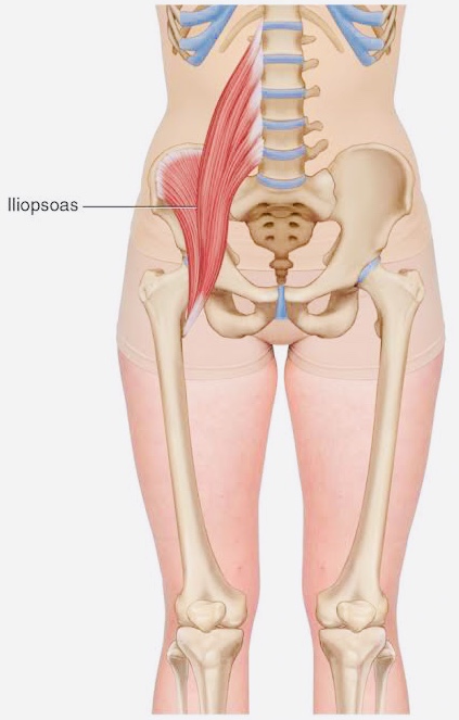
Surface anatomy of the lower extremity. In vertebrate anatomy, hip (or coxa in medical terminology) refers to either an anatomical region or a joint. 975 x 724 png 780 кб. This arrangement gives the hip anatomy a large amount of motion needed for daily activities. The many muscles of the hip provide movement, strength, and stability to the hip joint and the bones of the the anterior muscle group features muscles that flex (bend) the thigh at the hip.
The Key To Your Lower Back Pain Is In The Hips Try These 10 Stretches And Get Immediate Relief Live Love Fruit from livelovefruit.com The anatomy of the fascia lata and iliotibial tract. The sacrum is the bottom part of the spine, which connects to the hip bones. 400 x 400 jpeg 99 кб. When most people mention their back, what they are actually referring to is their spine. You just clipped your first slide! Clipping is a handy way to collect important slides you want to go back to later. Understanding how the different layers of the hip are built and connected can help you understand how the hip works, how it can be injured, and how challenging recovery can be when this joint is injured. Anatomy pictures of lower back and hip :
The muscles you probably know the best are your glutes (gluteal muscles), the large, strong muscles that attach to the back of your hip bones and comprise the buttocks.
Knowing the anatomy of your hip can help you understand the source of any hip pain. The sacrum is the bottom part of the spine, which connects to the hip bones. The hip joint is one of the most flexible joints in the entire human body. By dr arun pal singh. The bony pelvis protects the soft organs of pelvic cavity (bladder, lower colon, rectum, and reproductive organs). Surface anatomy of the lower extremity. This mri hip joint axial cross sectional anatomy tool is absolutely free to use. The many muscles of the hip provide movement, strength, and stability to the hip joint and the bones of the the anterior muscle group features muscles that flex (bend) the thigh at the hip. What makes up the pelvic girdle? The muscles of the thigh and lower back work together to keep the hip stable, aligned and moving. Want to learn more about it? Picture a man standing with the front of his pelvis tilting forward and his tailbone lifting. It also provides attachment points for many muscles that control the movements of the back.
Share :
Post a Comment
for "Anatomy Pictures Of Lower Back And Hip / Hip Sore Back Pain Try This Dr Sam Fitzgibbons Chiropractor"


Post a Comment for "Anatomy Pictures Of Lower Back And Hip / Hip Sore Back Pain Try This Dr Sam Fitzgibbons Chiropractor"